Corneal Topography Guide: Eye Surface Mapping for Vision Care
Introduction: Advanced Eye Mapping for Clearer Vision
When you look in the mirror, your eyes appear perfectly round. However, beneath that surface lies a complex landscape of microscopic curves that determine whether you’ll enjoy crystal-clear vision or struggle with blurry sight throughout your life.
Corneal topography is an advanced diagnostic technology that precisely maps this hidden terrain, providing eye care specialists with detailed information needed to optimize your vision. This comprehensive eye surface mapping creates a 3D blueprint of your cornea, revealing irregularities invisible to the naked eye.
At West Broward Eyecare Associates in Tamarac, we understand that your family’s vision needs extend beyond basic eye charts. Whether you’re concerned about your child’s changing prescription, considering LASIK surgery, or managing age-related eye conditions, corneal mapping provides the roadmap we need to deliver personalized care with confidence.
What Is Corneal Topography? (Quick Answer)
Corneal topography is a painless, non-invasive imaging test that creates detailed 3D maps of your cornea’s shape and curvature. This technology helps detect corneal irregularities early, plan precise vision correction procedures, and monitor eye health changes over time—all in under 10 minutes.
Key Benefits:
- Early Detection: Identifies eye conditions before symptoms appear
- Precision Planning: Guides LASIK, contact lens fitting, and cataract surgery
- Family Care: Safe for all ages, from children to seniors
- Preventive Health: Monitors changes to protect long-term vision
Understanding Corneal Topography: Advanced Eye Mapping Technology
What Is Corneal Mapping?
Corneal topography, also known as corneal mapping or photokeratoscopy, is a sophisticated imaging technology that captures thousands of measurement points across your cornea’s surface within seconds. Think of it as creating a detailed topographical map—but instead of mountains and valleys, we’re mapping the microscopic curves and corneal irregularities that affect your vision.
Your cornea is responsible for approximately 70% of your eye’s focusing power (about 43 diopters of the total 60 diopters), making its shape critical to clear vision. Even microscopic surface changes can significantly impact visual quality, which is why precise corneal mapping is essential for optimal eye care.
How Modern Corneal Topography Works
Today’s corneal topography systems use three primary technologies to create detailed eye surface maps:
1. Placido Disc Systems
These systems project concentric light rings onto your corneal surface and analyze reflection patterns. On healthy corneas, rings appear smooth and regular. Distortions indicate corneal irregularities that may affect vision quality.
Clinical Applications:
- Rapid screening for corneal diseases
- Contact lens fitting optimization
- Post-surgical monitoring
2. Scheimpflug Imaging Technology
This advanced corneal mapping technique uses rotating cameras to capture cross-sectional images from multiple angles. It creates comprehensive 3D maps of both front and back corneal surfaces.
Key Advantages:
- Complete corneal thickness analysis
- Detailed internal structure visualization
- Enhanced accuracy for complex cases
3. Scanning Slit Technology
Rapidly scanning light beams create precise measurements of corneal curvature and elevation. This technology excels at detecting subtle changes in corneal irregularities over time.
Primary Uses:
- Progressive disease monitoring
- Surgical planning enhancement
- Treatment effectiveness evaluation
Why Your Eye Doctor Recommends Corneal Topography
Early Detection of Corneal Irregularities
Many serious eye conditions begin with subtle changes invisible during routine examination. Corneal topography can detect early keratoconus and other ectatic conditions before symptoms appear, allowing for timely intervention that can preserve your vision for decades.
Key Conditions Detected:
- Keratoconus: Progressive thinning and bulging of the cornea
- Pellucid Marginal Degeneration: Inferior corneal thinning
- Irregular Astigmatism: Uneven corneal curvature
- Post-surgical Changes: Monitoring after previous eye procedures
Vision Correction Surgery Planning
Corneal topography is essential for determining eligibility for procedures such as LASIK, PRK, or refractive lens exchange. The detailed maps help your surgeon:
- Identify the safest treatment zones
- Customize ablation patterns for optimal results
- Screen out unsuitable candidates to prevent complications
- Plan toric lens implantation with precise axis alignment
Contact Lens Fitting Excellence
For complex prescriptions or specialty contact lenses, corneal topography provides the precise measurements needed for:
- Rigid gas-permeable lens fitting
- Scleral lens design for irregular corneas
- Orthokeratology (overnight vision correction)
- Post-surgical contact lens management
The Corneal Topography Experience: What to Expect
Before Your Appointment
Preparation Tips:
- Remove contact lenses 24-48 hours before testing (soft lenses) or up to 2 weeks (rigid lenses)
- Avoid eye makeup on the day of testing
- Bring current glasses and contact lens prescriptions
- List current medications and eye drops
During the Test
The Process:
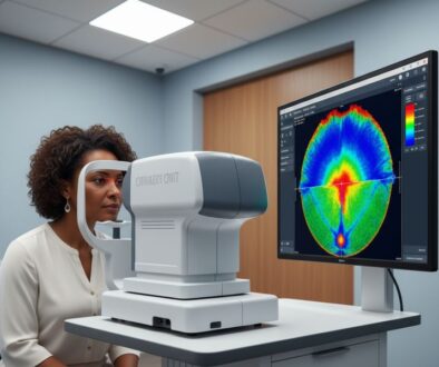
- Positioning: You’ll rest your chin and forehead against the device
- Fixation: Focus on a central light target while keeping your eye wide open
- Imaging: Multiple scans are captured in seconds – completely painless
- Blinking: Brief breaks between captures to maintain eye comfort
What You’ll Experience:
- Bright circular light patterns (similar to a camera flash)
- Brief periods of keeping your eyes open
- No physical contact with your eye
- Total test time: 5-10 minutes per eye
Understanding Your Results
Your corneal topography generates several types of maps:
Color-Coded Maps:
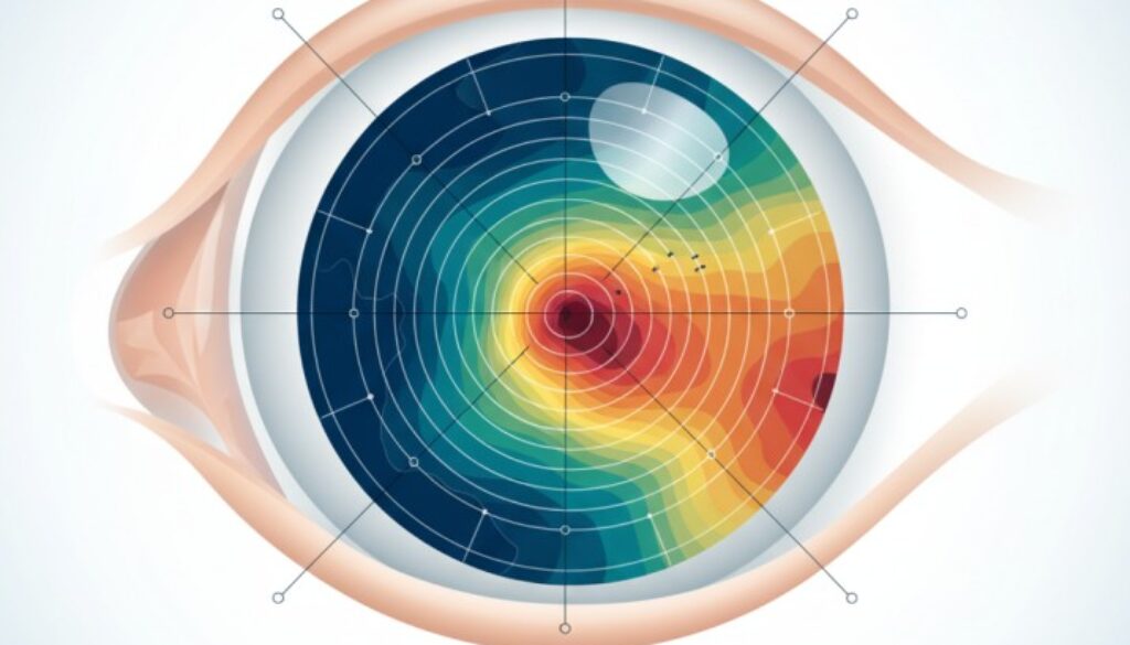
- Warm colors (red/orange): Steeper areas or higher elevation
- Cool colors (blue/purple): Flatter areas or lower elevation
- Green: Average curvature zones
Key Measurements:
- Keratometry values (K-readings): Corneal power in different meridians
- Astigmatism: Difference between steepest and flattest areas
- Irregularity indices: Mathematical measures of surface smoothness
Corneal Irregularities: Understanding Your Diagnosis
Regular vs. Irregular Astigmatism
Regular Astigmatism: Uniform steepening along a single corneal meridian that can be fully corrected with cylindrical lenses, typically resulting in 20/20 vision with proper correction.
Irregular Astigmatism: Nonuniform steepening that cannot be corrected by a cylindrical lens, often resulting in best-corrected vision of 20/50 or worse. This type requires specialized contact lenses or surgical intervention.
Keratoconus: The Progressive Challenge
Keratoconus typically manifests during puberty or early adulthood with symptoms such as blurry vision or a sudden decline in visual acuity. Key characteristics include:
Early Signs:
- Frequent prescription changes
- Difficulty achieving 20/20 vision with glasses
- Increased light sensitivity
- Ghost images or double vision
Advanced Features:
- Irregular corneal astigmatism and localized stromal thinning
- Steepening typically occurs in the inferotemporal region
- Progressive myopia and astigmatism
- Strong correlation between higher astigmatism levels and keratoconus diagnosis, with rates of 9.5% for 3.00D to <5.00D astigmatism and 17.4% for ≥5.00D astigmatism (2025 research data)
Treatment Options Based on Topography Findings
Mild Irregularities:
- Specialty soft contact lenses
- Custom glasses prescriptions
- Regular monitoring
Moderate Changes:
- Rigid gas-permeable contact lenses
- Corneal crosslinking to halt progression
- Intracorneal ring segments
Advanced Cases:
- Scleral contact lenses
- Corneal transplantation
- Combination therapies
Advanced Applications in Modern Eye Care
Artificial Intelligence and Machine Learning
Recent studies from 2020-2025 highlight the increasing role of machine learning in early detection and classification of keratoconus, with Random Forest algorithms achieving 96-98% accuracy in diagnosis.
AI-Enhanced Benefits:
- Earlier detection of subtle changes
- Improved risk stratification
- Personalized treatment recommendations
- Predictive modeling for disease progression
Multimodal Imaging Integration
Modern devices combine scanning-slit technology, Scheimpflug-based imaging, and anterior segment OCT to provide comprehensive corneal analysis, offering:
- Anterior and posterior surface mapping
- Corneal thickness measurements
- Biomechanical property assessment
- Corneal hysteresis evaluation as an early indicator of ectatic changes
Telemedicine and Remote Monitoring
Emerging technologies enable:
- Remote corneal topography screening
- AI-powered preliminary assessments
- Telehealth consultations with specialists
- Enhanced access to care in underserved areas
Market Growth and Technology Adoption
The global corneal topographers market reached $793-841 million in 2024 and is projected to grow to $890-984 million by 2025, with over 65% of eye care professionals relying on topography tools for pre-surgical assessments.
Corneal Topography for Families: Age-Specific Considerations
Pediatric Applications (Ages 6-17)
Why Children Need Corneal Topography:
- Early keratoconus detection during growth spurts
- Myopia management planning
- Sports vision optimization
- Pre-surgical evaluation for severe refractive errors
Making It Comfortable:
- Child-friendly explanations using familiar analogies
- Shorter test sessions with breaks
- Parent presence during testing
- Immediate results review to address concerns
Adult Applications (Ages 18-65)
Common Indications:
- Pre-LASIK/PRK evaluation
- Cataract surgery planning with premium IOLs
- Contact lens fitting optimization
- Occupational vision requirements
Senior Care (Ages 65+)
Age-Related Considerations:
- Corneal changes affecting cataract surgery
- Dry eye impact on measurements
- Medication effects on corneal health
- Medicare coverage considerations
Clinical Research and Evidence-Based Benefits
Recent Scientific Breakthroughs
2024 Research Highlights:
- Enhanced Keratoconus Detection. A 2024 study published in Scientific Reports demonstrated 98% training accuracy and 96% test accuracy using machine learning models for keratoconus detection in young adults aged 18-35.
- Biomechanical Integration: Incorporation of corneal hysteresis measurements with traditional topography improves sensitivity for detecting subclinical keratoconus and monitoring progression.
- Deep Learning Applications: Advanced U-Net neural networks now achieve corneal treatment zone segmentation in 0.06 seconds, facilitating rapid orthokeratology assessment.
Evidence-Based Treatment Outcomes
Surgical Success Rates:
- LASIK candidates properly screened: 98% satisfaction rates
- Corneal crosslinking with early topographic detection: 95% halting of progression
- Specialty contact lens success: 85% improved quality of life
Long-term Monitoring Benefits:
- 30% reduction in vision-threatening complications
- 40% improvement in early intervention success
- 60% better patient compliance with treatment
Insurance Coverage and Accessibility
When Insurance Typically Covers Corneal Topography
Diagnostic Coverage:
- Suspected keratoconus or corneal irregularities
- Pre-surgical evaluation for medically necessary procedures
- Post-surgical monitoring for complications
- Contact lens fitting for irregular corneas
Medicare Coverage (2025): Medicare covers corneal topography when medically necessary with proper documentation, with a standard reimbursement rate of $38.88.
Documentation Requirements:
- Clinical symptoms or risk factors
- Previous unsuccessful vision correction attempts
- Family history of corneal disease
- Specific medical indications
Financial Considerations
Typical Costs:
- Diagnostic corneal topography: $100-$250 (standalone test)
- Contact lens evaluation, including topography: $150-$400
- Serial monitoring: $100-$200 per follow-up visit
- Combined with other tests, Package pricing is typically available
Payment Options:
- Insurance benefits verification
- Flexible spending accounts (FSA/HSA)
- Payment plan arrangements
- Package deals for comprehensive care
Choosing the Right Provider: What to Look For
Technology and Expertise
Advanced Equipment:
- Latest generation topography systems
- Multimodal imaging capabilities
- AI-assisted analysis tools
- Regular calibration and maintenance
Provider Qualifications:
- Board certification in ophthalmology
- Specialty training in corneal diseases
- Experience with complex cases
- Continuing education in the latest techniques
Patient-Centered Care Approach
What Sets Us Apart at West Broward Eyecare Associates:
Family-Focused Philosophy: We understand that vision care affects the entire family. Our approach considers:
- Multi-generational eye health patterns
- Lifestyle and educational needs
- Long-term vision preservation goals
- Family budget and insurance considerations
Comprehensive Care Integration:
- Seamless coordination between general and specialty care
- Educational resources for all family members
- Emergency and urgent care availability
- Community partnerships with schools and organizations
Technology with a Human Touch:
- State-of-the-art diagnostic equipment
- Warm, experienced technicians
- Clear explanations of results
- Collaborative treatment planning
Preparing for Your Corneal Topography Appointment
Before You Arrive
Contact Lens Wearers:
- Soft lenses: Remove 24-48 hours before testing
- Toric soft lenses: Remove 1 week before
- Rigid gas-permeable lenses: Remove 2-4 weeks before
- Extended wear lenses: Consult our office for specific timing
Day of Testing:
- Avoid eye makeup and creams
- Bring current glasses and contact lenses
- List all medications and eye drops
- Arrive 15 minutes early for paperwork
Questions to Ask Your Eye Doctor
About Your Results:
- What do my corneal topography measurements indicate?
- How do these results compare to normal ranges?
- Are there any areas of concern requiring monitoring?
- What treatment options are available if needed?
About Follow-up:
- How often should I have repeat testing?
- What changes should I watch for?
- How will this affect my vision correction options?
- Should family members be screened?
The Future of Corneal Topography
Emerging Technologies
Next-Generation Developments:
- Real-time AI analysis during testing
- Integration with electronic health records
- Portable devices for community screening
- AI-powered topography systems and multimodal imaging technologies are becoming indispensable tools for precision eye care
Personalized Vision Care
Precision Medicine Applications:
- Genetic testing integration
- Customized treatment algorithms
- Predictive modeling for outcomes
- Population health management
Enhanced Patient Experience:
- Faster testing protocols
- Improved comfort during procedures
- Interactive result explanations
- Mobile app integration for tracking
Take Action for Your Vision Health
When to Schedule Corneal Topography
Immediate Evaluation Needed:
- Sudden vision changes
- Difficulty with current glasses or contacts
- Family history of keratoconus
- Considering vision correction surgery
Routine Monitoring:
- Annual screening for at-risk individuals
- Pre-surgical evaluation
- Contact lens fitting optimization
- Sports vision enhancement
Your Next Steps
Getting Started:
- Schedule a Comprehensive Eye Exam: Call West Broward Eyecare Associates to discuss your vision concerns.
- Insurance Verification: We’ll help determine your coverage benefits
- Preparation: Follow pre-test instructions for optimal results
- Follow-up Planning: Develop a personalized monitoring schedule
Medical References and Sources:
- American Academy of Ophthalmology – Corneal Topography Guidelines
- National Center for Biotechnology Information – Corneal Topography Review
- StatPearls – Corneal Topography Clinical Applications
- Review of Ophthalmology – Corneal Topography Advances
- Scientific Reports – AI in Keratoconus Detection
Conclusion: Your Vision Health Starts with Precise Mapping
Corneal topography represents one of the most significant advances in modern eye care, transforming how we detect, diagnose, and treat vision-threatening conditions. This sophisticated corneal mapping technology provides detailed insights that were impossible just decades ago, enabling earlier detection and more precise treatment of corneal irregularities.
Whether you’re managing a complex eye condition, planning vision correction surgery, or ensuring optimal eye health for your family, corneal topography offers the precision and accuracy needed for confident decision-making. At West Broward Eyecare Associates, we combine cutting-edge corneal mapping technology with personalized care your family deserves.
Why Choose Advanced Corneal Mapping?
Our commitment to excellence, comprehensive approach, and community focus ensures you receive not just advanced corneal topography diagnostics, but ongoing support needed to maintain healthy vision throughout your lifetime. From early corneal irregularities detection to post-surgical monitoring, we’re your partners in vision health.
Take Action Today: Don’t wait for symptoms to appear. Early detection through corneal topography can preserve and enhance your vision for years to come. Contact West Broward Eyecare Associates today to discover how this remarkable technology can benefit you and your family.
Schedule Your Corneal Mapping Consultation 📞 Visit Our📍 Location: Tamarac, F.L
About West Broward Eyecare Associates: Our experienced eye care professionals have served Tamarac and surrounding communities for over years, providing comprehensive vision care with the latest corneal topography technology. Our commitment to family-centered care and clinical excellence makes us the trusted choice for protecting and enhancing vision health for patients of all ages.
FAQs
-
A corneal topography test takes 5-10 minutes for both eyes. The actual scanning process takes only seconds per eye, with brief breaks between captures for blinking and comfort.

