

Optomap Eye Exam: Comprehensive Retinal Screening in Tamarac
Introduction
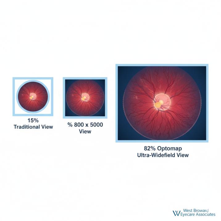
When it comes to protecting your family’s vision, early detection makes all the difference. Advanced retinal imaging technology can now document up to 82% of your retina in a single capture lasting less than half a second—without the need for uncomfortable dilating eye drops.
Optomap retinal imaging has revolutionized how eye care professionals assess retinal health, offering a comfortable, comprehensive alternative to traditional dilated eye examinations. This ultra-widefield technology enables the detection of eye diseases often before symptoms appear, potentially saving your sight and improving treatment outcomes.
Whether you’re a busy parent coordinating family healthcare, a senior managing multiple health conditions, or someone seeking the most advanced eye care available, understanding Optomap technology empowers you to make informed decisions about your vision health.
What Is Optomap Retinal Imaging?
Optomap retinal imaging is an advanced diagnostic technology that captures ultra-widefield images of your retina without requiring pupil dilation. Developed by Optos, this revolutionary system provides eye care professionals with unprecedented views of retinal structures in just a quarter of a second.
Key Technical Specifications:
- Coverage: Up to 82% (200 degrees) of the retina in a single capture
- Enhanced Coverage: Up to 97% (220 degrees) with auto-montage technology
- Capture Time: Less than 0.25 seconds per eye
- Patient Comfort: No dilation drops required
- Image Quality: High-resolution, permanent digital records
Did You Know? Traditional retinal imaging methods typically show only 10-15% of your retina at one time, while Optomap captures more than 80% in a single image.
How Optomap Technology Works
Multi-Spectral Laser Technology
Unlike conventional devices that use full-spectrum white light, Optomap technology incorporates low-powered laser wavelengths that scan simultaneously. Each laser wavelength examines different retinal layers:
- Green Laser (532 nm): Scans the sensory retina to the pigment epithelial layers
- Red Laser (633 nm): Examines deeper RPE to choroidal structures
- Blue Laser (488 nm): Enables fluorescein angiography and color RGB imaging
- Infrared Laser (802 nm): Facilitates specialized angiography procedures
This multi-spectral approach allows comprehensive examination of retinal substructures that would otherwise require multiple separate tests.
The Patient Experience
Optomap examinations are remarkably simple and comfortable:
- Positioning: You simply look into the device and focus on a small LED target
- Imaging: A brief, painless flash captures the retinal image in 0.25 seconds
- Review: Your eye care professional immediately reviews results with you
- No Recovery: Resume normal activities immediately—no vision impairment
Key Benefits vs Traditional Dilation
Comfort and Convenience Advantages
| Optomap Imaging | Traditional Dilation |
|---|---|
| ✅ No eye drops required | ❌ Stinging drops necessary |
| ✅ Immediate normal vision | ❌ 4-6 hours blurred vision |
| ✅ 0.25-second capture | ❌ 30+ minute process |
| ✅ Drive immediately after | ❌ Transportation needed |
| ✅ Return to work/activities | ❌ Limited functionality |
Superior Detection Capabilities
Clinical research demonstrates significant advantages:
- 30% improvement in retinal lesion detection when combined with traditional exams
- 19% more diabetic retinopathy cases detected compared to standard photography
- 15% of cases received higher severity grades, enabling earlier intervention
Comprehensive Disease Detection
Optomap retinal imaging excels at detecting:
Eye Conditions:
- Diabetic retinopathy (early-stage detection)
- Age-related macular degeneration
- Retinal detachments and tears
- Glaucoma-related retinal changes
- Blood vessel abnormalities
Systemic Health Indicators: Since your retina contains the only blood vessels visible directly in the body, signs of systemic diseases often appear here first:
- Cardiovascular disease
- Hypertension complications
- Diabetes-related changes
- Stroke risk indicators
- Certain cancers
Clinical Evidence and Research
Peer-Reviewed Scientific Studies
Enhanced Diabetic Retinopathy Detection (2024) Published Research: PMC Database
A landmark study analyzing 266 retinal images found that Optomap ultra-widefield imaging identifies 19% more diabetic retinopathy cases compared to traditional seven-field photography. Significantly, 15% of cases received higher severity grades when the full 200-degree view was utilized, potentially leading to earlier intervention and better patient outcomes.
30% Improvement in Lesion Detection Source: Eye and Brain Journal
Research from the New England College of Optometry revealed that combining retinal imaging with traditional examination increased overall retinal lesion detection by 30% compared to ophthalmoscopy alone. In cases of disagreement between methods, adjudicators agreed with the image-assisted approach in over 70% of instances.
Ultra-Widefield Clinical Validation Published Research: PMC Database
Current research confirms modern Optomap systems capture 82% of the retina in standard mode, with auto-montage capabilities extending coverage to 97%. Studies indicate that 66% of retinal pathology occurs beyond the reach of standard fundus cameras, supporting the clinical value of wide-field imaging.
Clinical Trial Evidence
With more than 2,500 published and ongoing clinical trials, plus thousands of case studies and testimonials, research consistently demonstrates the long-term value of Optomap imaging in:
- Early disease diagnosis
- Treatment planning optimization
- Patient education and engagement
- Long-term health monitoring
Cost and Insurance Coverage
Investment in Advanced Eye Care
Current Pricing (2025):
- Typical Range: $29-$65 per examination
- Most Common: $30-$45 at participating practices
- Regional Variations: Pricing varies by location and imaging complexity
Insurance Coverage Guidelines
Vision Insurance:
- Generally not covered for routine screening
- Some plans cover when medically indicated
- FSA/HSA funds may be applicable
Medical Insurance:
- May be covered when medically necessary
- Diabetes monitoring is often covered
- Specific eye conditions typically qualify
- Check with your provider for coverage details
Value Assessment
Many patients find the investment worthwhile, considering:
- Time savings: No recovery period from dilation
- Convenience: Immediate return to normal activities
- Superior documentation: Permanent digital health records
- Enhanced detection: Earlier disease identification
- Family-friendly: Comfortable for all ages
Who Should Consider Optomap?
Ideal Candidates for Optomap Imaging
Routine Eye Care:
- Annual comprehensive eye examinations
- Adults seeking preventive care
- Patients want detailed documentation
- Those preferring non-invasive procedures
Specific Medical Conditions:
- Diabetes: Enhanced diabetic retinopathy monitoring
- Hypertension: Blood vessel health assessment
- Family History: Genetic eye disease screening
- High Myopia: Increased retinal detachment risk
Lifestyle Considerations:
- Busy Professionals: Cannot afford vision downtime
- Parents: Managing multiple family appointments
- Seniors: Avoiding medication interactions
- Active Adults: Immediate return to activities required
Special Population Benefits
Pediatric Eye Care: Optomap is particularly valuable for children because:
- No eye drops reduce anxiety and resistance
- Quick procedure maintains attention span
- Superior peripheral view detects childhood conditions
- Creates baseline records for future comparison
Senior Adults: Benefits for older patients include:
- No medication interactions with dilating drops
- Immediate driving capability after the appointment
- Enhanced monitoring of age-related conditions
- Clear documentation for coordinated care
Making the Right Choice: Optomap vs Dilation
When Optomap Is Preferred
Optimal Scenarios:
- Routine annual eye examinations
- Diabetic retinopathy screening
- Patient sensitivity to eye drops
- Scheduling constraints requiring efficiency
- Pediatric eye examinations
- Documentation for insurance or legal purposes
When Traditional Dilation May Still Be Necessary
Professional medical organizations, including the American Academy of Ophthalmology and American Optometric Association, still recommend:
- First comprehensive eye examination
- Emergency eye conditions (flashes, floaters, sudden vision changes)
- Complex retinal pathology investigation
- Detailed optic nerve assessment for glaucoma
- Annual dilated exams for diabetic patients
Integrated Approach
Leading eye care professionals increasingly recommend combining both technologies when appropriate, leveraging:
- Optomap’s superior field of view and documentation
- Dilation’s detailed stereoscopic examination capabilities
- Patient comfort with comprehensive diagnostic accuracy
Resources and Scientific Citations
This article is based on peer-reviewed research, clinical studies, and authoritative sources in ophthalmology and retinal imaging. The following key resources were consulted to ensure accuracy and provide evidence-based information:
1. Enhanced Diabetic Retinopathy Detection Study
“Optomap ultrawide field imaging identifies additional retinal abnormalities in patients with diabetic retinopathy”
Source: PMC – National Center for Biotechnology Information
https://pmc.ncbi.nlm.nih.gov/articles/PMC4376301/
This landmark study demonstrates that Optomap ultra-widefield imaging identifies 19% more diabetic retinopathy cases compared to traditional seven-field photography. The research involved 266 images and found that 15% of cases received higher severity grades when using the full 200-degree view, supporting earlier intervention and better patient outcomes.
2. Clinical Validation of Image-Assisted Retinal Examination
“Image-assisted fundus examination may enhance detection of retinal lesions by 30%”
Source: Eye and Brain Journal – Peer-reviewed Clinical Research
https://www.roswelleyeclinic.com/technology/optomap/
Published research from the New England College of Optometry revealed that combining retinal imaging with traditional examination increased overall retinal lesion detection by 30% compared to ophthalmoscopy alone. The study found good agreement between image-assisted and traditional fundus examination, with adjudicators agreeing with the image-assisted method in over 70% of cases where there was disagreement.
3. Ultra-Widefield Retinal Imaging: Clinical Advances and Applications
“Ultra-widefield retinal imaging: an update on recent advances”
Source: PMC – National Center for Biotechnology Information
https://pmc.ncbi.nlm.nih.gov/articles/PMC6971964/
This comprehensive review examines the importance of ultra-widefield imaging in modern ophthalmology, detailing the technical capabilities of Optomap technology, including its ability to capture 82% of the retina in a single image and up to 97% with auto-montage functionality. The research emphasizes the clinical significance of peripheral retinal visualization for early disease detection.
Conclusion
Optomap retinal imaging represents a significant advancement in eye care technology, offering comprehensive retinal assessment without the inconvenience and discomfort of traditional dilation. With its ability to capture up to 82% of the retina in a single, comfortable examination, this technology has revolutionized how eye care professionals detect, monitor, and treat eye diseases.
The scientific evidence supporting Optomap’s diagnostic capabilities is substantial, with studies demonstrating improved detection rates for various retinal conditions. Whether you’re managing a chronic condition like diabetes, seeking routine preventive care, or concerned about a family history of eye disease, Optomap technology provides valuable insights into your eye health.
Key takeaways for patients considering Optomap imaging:
- Superior coverage: 82% retinal visualization vs. 15% traditional methods
- Enhanced detection: 30% improvement in identifying retinal lesions
- Patient comfort: No dilation drops, immediate normal vision
- Comprehensive documentation: Permanent digital records for monitoring
- Family-friendly: Safe and comfortable for all ages
As with any medical decision, the choice between Optomap imaging and traditional dilation should be made in consultation with your qualified eye care professional. By understanding the benefits, limitations, and appropriate applications of each approach, you can make informed decisions that align with your health needs, lifestyle, and preferences.
Regular comprehensive eye examinations remain essential for maintaining optimal vision health throughout your life. Early detection and appropriate treatment of eye diseases can preserve your sight and enhance your quality of life for years to come. The investment in advanced diagnostic technology like Optomap represents an investment in your long-term vision health.
When combined with the expertise of skilled eye care professionals and your commitment to regular preventive care, this powerful technology helps ensure that you and your family can continue to see the world clearly.
This article provides educational information about Optomap retinal imaging and should not replace professional medical consultation. Consult with qualified eye care professionals for personalized examination recommendations and treatment decisions.
About West Broward Eyecare Associates: As your trusted vision guardians in Tamarac, we combine cutting-edge eye care technology with the personal attention your family deserves. From your child’s first eye exam to managing complex conditions with advanced technology, we’re here to protect and preserve the gift of sight for every stage of life.
FAQs
-
Optomap is a digital scanning technology that captures ultra-wide images of your retina using laser light. The scan takes less than one second per eye and requires no eye drops or physical contact with your eye.


