

Optical Coherence Tomography for Clearer Vision
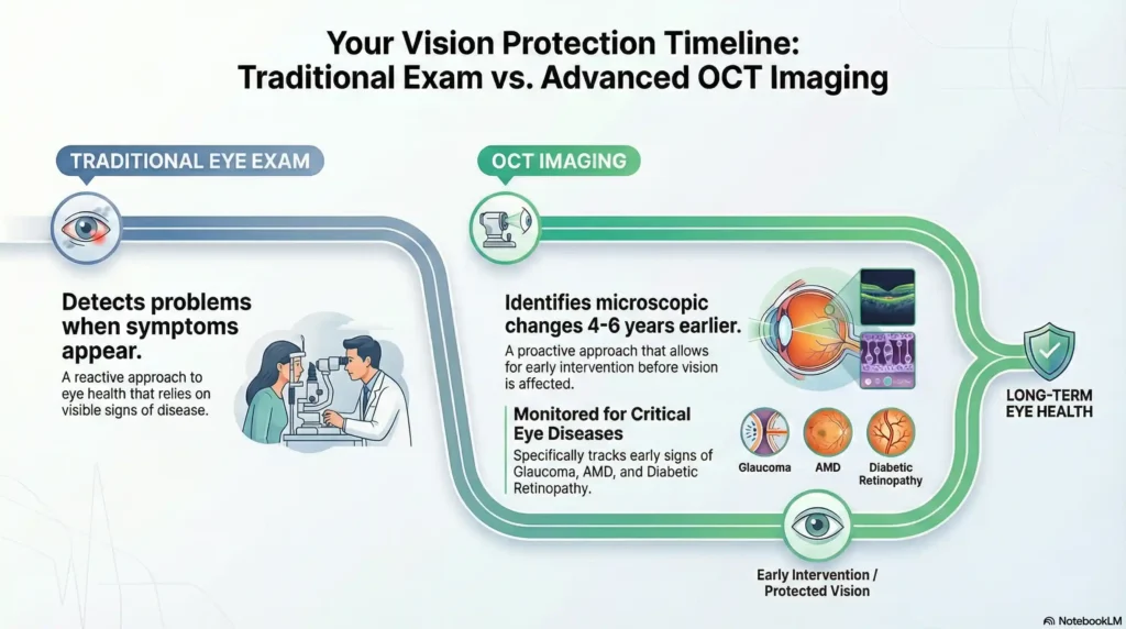
Optical Coherence Tomography (OCT) is a non-invasive imaging test that uses light waves to take cross-section pictures of your retina, allowing for the detection of vision-threatening diseases like glaucoma and AMD up to 4–6 years before symptoms appear. At West Broward Eyecare, we use this advanced technology to provide families in Tamarac and West Broward County with a precise, microscopic look at their eye health to prevent permanent vision loss.
[Protect Your Vision – Schedule Your OCT Scan Today]
The Technology That Sees Beyond the Surface
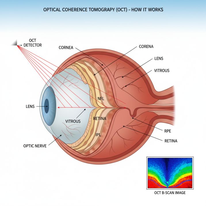
Think of an OCT scan for eyes as creating a detailed “slice” through your retina, similar to how an MRI creates images of your brain. However, instead of using magnetic fields, this advanced eye imaging employs near-infrared light to safely penetrate eye tissues and capture microscopic details invisible to traditional examination methods.
Key benefits of OCT imaging include:
- Early detection: Identifies disease 4-6 years before symptoms develop
- Precise monitoring: Tracks changes with micrometer accuracy
- Non-invasive process: Safe, comfortable, and quick examination
- Comprehensive assessment: Evaluates all retinal layers simultaneously
Why OCT Matters for Your Vision Health
The transformative power of optical coherence tomography lies in its ability to reveal the earliest signs of eye disease when treatment is most effective. Many conditions that threaten vision—including glaucoma, often called the “silent thief of sight“—develop without noticeable symptoms until significant damage has already occurred.
OCT technology changes this paradigm by providing detailed structural information that enables proactive treatment, often preventing vision loss entirely.
How OCT Works: The Science Behind the Technology
Understanding how OCT works helps appreciate why this technology has become indispensable in modern eye care. The process relies on a principle called low-coherence interferometry, which sounds complex but operates on a simple concept similar to echolocation.
The Light Wave Process
OCT scanners emit a carefully controlled beam of near-infrared light into your eye. This light safely penetrates your eye tissues and reflects off different structures at varying depths. Just as radar measures distance by timing how long radio waves take to bounce back, OCT measures the time it takes for light waves to return from different layers of your eye.
The key innovation lies in OCT’s ability to measure these incredibly tiny time differences – we’re talking about measurements accurate to within a few micrometers (that’s about 1/50th the width of a human hair). This precision allows the system to create detailed cross-sectional images showing each retinal layer with extraordinary clarity.
Creating the Image
The reflected light waves are compared to a reference beam using sophisticated computer algorithms. When these light waves interfere with each other, they create patterns that the OCT system interprets to build a detailed map of your eye’s internal structures. This process happens in real-time, allowing your eye doctor to see live images of your retina during the scan.
Modern OCT systems can capture up to 80,000 measurements per second, enabling comprehensive imaging of your entire retina in just seconds. This speed ensures comfort during the examination while providing your doctor with incredibly detailed information about your eye health.
Types of OCT Technology
OCT technology has evolved significantly since its introduction, with each advancement offering improved image quality and diagnostic capabilities. Understanding these different types helps explain why some OCT systems provide superior imaging for specific conditions.
Time-Domain OCT (TD-OCT)
The original OCT technology, time-domain systems, were groundbreaking when introduced, but are now largely superseded by newer technologies. TD-OCT systems provided good images but were relatively slow, taking several seconds to capture each scan line.
Spectral-Domain OCT (SD-OCT)
Currently, the most widely used OCT technology, spectral-domain systems, offer significant improvements over time-domain imaging. SD-OCT provides:
- Faster scanning speeds: Complete retinal scans in seconds rather than minutes
- Higher resolution: Better visualization of subtle retinal changes
- Improved patient comfort: Reduced examination time and movement artifacts
- Enhanced disease detection: Superior ability to identify early pathological changes
Swept-Source OCT (SS-OCT)
The newest generation of OCT technology, swept-source systems, represents the cutting edge of retinal imaging. SS-OCT offers several advantages:
- Deeper tissue penetration: Better visualization of structures beneath the retina
- Faster scanning: Even quicker image acquisition than SD-OCT
- Reduced artifacts: Less interference from cataracts or other media opacities
- Wider imaging areas: Ability to capture larger retinal areas in a single scan
OCT Angiography (OCTA)
A revolutionary extension of traditional OCT, OCT angiography provides detailed images of blood vessels in your retina without requiring dye injection. This technology detects blood flow by analyzing subtle changes in light reflection over time, creating detailed maps of your retinal circulation.
OCTA is particularly valuable for detecting:
- Diabetic retinopathy changes
- Macular degeneration complications
- Glaucoma-related circulation problems
- Various retinal vascular disorders
Eye Diseases Detected by OCT Scans
OCT imaging excels at detecting and monitoring a wide range of eye conditions, often identifying problems before they cause noticeable symptoms. This early detection capability can be vision-saving, allowing for prompt treatment that prevents or slows disease progression.
Glaucoma Detection and Monitoring
Glaucoma, often called the “silent thief of sight,” typically causes no symptoms until significant vision loss has occurred. OCT technology revolutionizes glaucoma detection by measuring the thickness of your retinal nerve fiber layer and ganglion cell complex – areas that show thinning before vision changes become apparent.
Research shows that OCT can detect glaucomatous damage up to 4-6 years earlier than traditional methods. For patients with a family history of glaucoma or other risk factors, this early detection can be sight-saving.
OCT advantages for glaucoma include:
- Precise measurement of nerve fiber layer thickness
- Detection of subtle progressive changes over time
- Assessment of optic nerve head structure
- Monitoring treatment effectiveness
Age-Related Macular Degeneration (AMD)
AMD affects central vision and becomes more common with age. OCT provides a detailed visualization of the macula, allowing doctors to:
- Detect drusen (yellowish deposits under the retina)
- Identify retinal pigment epithelium changes
- Monitor for wet AMD complications
- Track disease progression over time
- Guide treatment decisions for advanced cases
The ability to see fluid accumulation or abnormal blood vessel growth beneath the retina makes OCT invaluable for AMD management.
Diabetic Retinopathy and Macular Edema
For patients with diabetes, OCT serves as a crucial monitoring tool. The technology excels at detecting:
- Diabetic macular edema: Fluid accumulation in the central retina
- Retinal thickening: Early signs of diabetic damage
- Microaneurysms and hemorrhages: Small blood vessel abnormalities
- Treatment response: Monitoring improvement following therapy
OCT’s ability to precisely measure retinal thickness helps doctors determine when treatment is needed and assess its effectiveness.
Epiretinal Membranes
These thin sheets of scar tissue that can develop on the retinal surface often cause vision distortion. OCT clearly shows:
- Membrane location and thickness
- Retinal distortion patterns
- Need for surgical intervention
- Post-surgical healing progress
Macular Holes
Full-thickness defects in the central retina, macular holes, can severely impact central vision. OCT provides:
- Precise hole size measurements
- Assessment of surrounding retinal changes
- Surgical planning information
- Monitoring of healing after treatment
Central Serous Chorioretinopathy
This condition involves fluid accumulation beneath the retina, often affecting young to middle-aged adults. OCT reveals:
- Extent of subretinal fluid
- Retinal pigment epithelium changes
- Disease activity levels
- Treatment response monitoring
Retinal Vein and Artery Occlusions
When blood vessels in the retina become blocked, OCT helps assess:
- Retinal swelling extent
- Macular involvement
- Treatment planning needs
- Recovery progress monitoring
The OCT Examination Experience
Understanding what to expect during an OCT scan helps patients feel comfortable and ensures optimal results. The examination is quick, painless, and requires no special preparation in most cases.
Before Your OCT Scan
No special preparation is typically needed, though your eye doctor may recommend:
- Arriving with current glasses or contact lenses
- Bring a list of current medications
- Arranging transportation if pupil dilation is planned
- Allowing extra time for a comprehensive evaluation
During the Examination
The OCT scanning process is straightforward and comfortable:
- Positioning: You’ll sit comfortably in front of the OCT machine and rest your chin on a support platform
- Focusing: Look at a small fixation target inside the machine (usually a green light or simple image)
- Scanning: The machine captures images automatically while you maintain steady fixation
- Duration: Each eye typically takes 1-2 minutes to scan completely
- Comfort: Nothing touches your eye, and you’ll only see harmless light patterns
Patient Experience Tips
To ensure the best possible images:
- Stay relaxed: The procedure is completely painless
- Maintain steady focus: Keep looking at the target light even if you see scanning beams
- Breathe normally: Don’t hold your breath during scanning
- Ask questions: Your technician can explain what’s happening throughout the process
Cost and Insurance Considerations
OCT pricing in 2025: As an additional diagnostic test, OCT scans typically cost between $39-$99 beyond the standard eye examination fee, with pricing varying by practice location and technology used.
Insurance coverage: Medicare and many private insurance plans cover OCT when medically necessary for diagnosed conditions such as glaucoma, macular degeneration, or diabetic retinopathy. For routine screening in healthy individuals, OCT is often considered an elective upgrade with out-of-pocket costs.
Value consideration: The early detection capabilities and comprehensive health information provided by OCT often justify the additional cost, particularly for patients over 40 or those with risk factors for eye disease.
After the Scan
Immediate results: Your doctor can review images immediately after scanning, often discussing findings during the same visit. If pupil dilation is used, you may experience:
- Light sensitivity for 2-4 hours
- Blurred near vision temporarily
- Need for sunglasses outdoors
Most patients can resume normal activities immediately after OCT scanning, though driving may be delayed if pupils were dilated.
Benefits of OCT for Early Disease Detection
The transformative power of OCT lies in its ability to detect eye diseases in their earliest stages, often years before symptoms develop. This early detection capability offers numerous advantages for preserving vision and maintaining quality of life.
Earlier Detection Than Traditional Methods
Traditional eye examinations, while valuable, have limitations in detecting subtle retinal changes. OCT overcomes these limitations by:
- Revealing microscopic changes: Detecting alterations as small as a few micrometers
- Quantifying progression: Providing precise measurements for tracking disease advancement
- Identifying risk factors: Spotting early warning signs before irreversible damage occurs
- Enabling proactive treatment: Allowing intervention before symptoms appear
Research demonstrates that OCT can detect certain conditions 3-4 years earlier than conventional examination methods, providing a crucial window for preventive treatment.
Precise Measurement and Monitoring
OCT’s quantitative capabilities transform eye care from subjective assessment to objective measurement:
- Thickness measurements: Precise tracking of retinal layer changes over time
- Volume calculations: Three-dimensional assessment of retinal structures
- Progression analysis: Mathematical determination of disease advancement rates
- Treatment response: Objective measurement of therapy effectiveness
Non-Invasive and Safe
Unlike some traditional diagnostic tests, OCT offers several safety advantages:
- No radiation exposure: Uses harmless near-infrared light
- No contrast injections: Provides detailed images without dyes or medications
- No contact with eyes: Maintains complete comfort during examination
- Repeatable safely: Can be performed as often as needed without risk
Comprehensive Retinal Assessment
Modern OCT systems provide a holistic evaluation of retinal health:
- Multiple-layer analysis: Simultaneous assessment of all retinal layers
- Vascular imaging: Blood vessel evaluation through OCT angiography
- Structural assessment: Detailed visualization of retinal architecture
- Functional correlation: Linking structural changes to visual function
OCT vs. Traditional Eye Examination Methods
Understanding how OCT compares to other eye examination techniques helps appreciate its unique value in comprehensive eye care. While traditional methods remain important, OCT provides capabilities that significantly enhance diagnostic accuracy and monitoring precision.
OCT vs. Direct Ophthalmoscopy
Traditional ophthalmoscopy involves using a handheld light and a magnifying lens to examine the retina directly. While this method provides valuable information, it has limitations:
- Limited magnification: Difficulty seeing subtle changes
- Subjective interpretation: Results depend on the examiner’s experience
- No permanent record: Cannot easily track changes over time
- 2D visualization only: Limited depth information
OCT advantages:
- High magnification: Microscopic detail visualization
- Objective measurements: Precise, numerical data
- Permanent documentation: Digital storage for comparison
- 3D cross-sectional views: Complete structural assessment
OCT vs. Fundus Photography
Traditional fundus photography captures color images of the retina surface, providing excellent documentation of visible changes. However, it cannot:
- See beneath the retinal surface
- Measure retinal thickness accurately
- Detect early structural changes
- Provide quantitative progression data
OCT complements fundus photography by adding:
- Subsurface structural visualization
- Precise thickness measurements
- Early disease detection capabilities
- Quantitative monitoring tools
OCT vs. Fluorescein Angiography
Fluorescein angiography involves injecting dye to visualize retinal blood vessels. While valuable for certain conditions, it requires:
- Intravenous injection with potential allergic reactions
- Multiple images over 10-15 minutes
- Possible side effects (nausea, skin discoloration)
- Patient preparation and recovery time
OCT angiography provides:
- No injection requirements
- Rapid image acquisition
- No adverse reactions
- Immediate results
Integrated Approach
The most effective eye care combines OCT with traditional examination methods:
- Clinical examination: Overall eye health assessment
- Visual field testing: Functional vision evaluation
- OCT imaging: Structural analysis and monitoring
- Additional tests: As needed for specific conditions
This comprehensive approach ensures both current eye health evaluation and future vision protection.
Latest Advances in OCT Technology
OCT technology continues evolving rapidly, with innovations enhancing image quality, expanding diagnostic capabilities, and improving patient accessibility. These advances promise even better eye care outcomes and more convenient examination experiences.
Artificial Intelligence Integration
AI-powered OCT analysis represents a major advancement in eye care diagnostics:
Automated disease detection: AI algorithms can identify pathological changes with accuracy matching or exceeding human specialists. These systems analyze thousands of image features simultaneously, detecting subtle patterns that might be missed by visual inspection alone.
Predictive analytics: Advanced AI models can predict disease progression risk by analyzing current OCT images alongside patient history. This capability helps doctors make more informed treatment decisions and provides patients with better prognostic information.
Quality assurance: AI systems automatically assess image quality, ensuring optimal diagnostic value and identifying when rescanning might be beneficial.
Treatment planning: Machine learning algorithms help optimize treatment protocols by analyzing response patterns from similar cases, personalizing care for better outcomes.
Portable and Home-Based OCT Systems
Revolutionary developments in OCT miniaturization are making this technology more accessible:
Community-based screening: Portable OCT units enable screening programs in underserved areas, bringing advanced eye care to communities with limited access to specialists.
Home monitoring systems: Emerging technologies allow patients with chronic conditions to perform OCT scans at home, enabling more frequent monitoring without office visits. This is particularly valuable for conditions requiring regular assessment, such as macular degeneration or diabetic retinopathy.
Telemedicine integration: Remote OCT interpretation allows specialists to provide expert analysis regardless of geographic location, expanding access to specialized care.
Enhanced Imaging Capabilities
Wider field imaging: Newer OCT systems capture larger retinal areas in single scans, providing more comprehensive assessment and reducing examination time.
Higher resolution: Improved light sources and detection systems offer even finer detail visualization, enabling detection of increasingly subtle pathological changes.
Faster scanning: Advanced systems complete comprehensive retinal imaging in seconds, improving patient comfort and reducing motion artifacts.
Multi-modal imaging: Integration of OCT with other imaging modalities provides complementary information for a more complete diagnostic assessment.
Swept-Source Technology Advances
The latest swept-source OCT systems offer significant improvements:
Deeper penetration: Enhanced ability to image through cataracts and other media opacities, extending OCT utility to more patients.
Choroidal imaging: Better visualization of blood vessels and structures beneath the retina, important for conditions like macular degeneration.
Reduced artifacts: Minimized interference from eye movements and other factors that can degrade image quality.
Faster acquisition: Extremely rapid imaging enables real-time visualization and improved patient tolerance.
Who Should Get OCT Scans?
OCT imaging benefits virtually everyone concerned about maintaining healthy vision, from those seeking baseline documentation to individuals requiring monitoring of existing eye conditions. Understanding current recommendations helps patients make informed decisions about comprehensive eye care.
Universal Screening Guidelines for 2025
Adults Age 40 and Over. Age-related changes make the OCT scan for the eyes valuable for establishing baseline measurements and detecting early disease indicators. The American Academy of Ophthalmology recommends comprehensive eye examinations every 2-4 years for adults in this age group, with OCT providing enhanced diagnostic capabilities.
Family History Considerations. Individuals with relatives who have experienced glaucoma, macular degeneration, or other hereditary eye conditions benefit significantly from earlier and more frequent OCT monitoring, as genetics play a substantial role in eye disease development.
High-Priority Groups Requiring Regular OCT Monitoring
Diabetes Patients (Annual OCT Required) All individuals with Type 1 or Type 2 diabetes should receive annual optical coherence tomography examinations to monitor for:
- Diabetic retinopathy progression
- Macular edema development
- Retinal blood vessel changes
- Treatment response assessment
Glaucoma Risk Factors: People with the following characteristics require regular OCT monitoring:
- Elevated eye pressure (over 21 mmHg)
- Family history of glaucoma
- African American heritage (40+ years)
- Hispanic heritage (60+ years)
- Severe nearsightedness (high myopia)
Cardiovascular Disease and Hypertension: These systemic conditions affect retinal blood vessels, making OCT monitoring valuable for detecting circulation-related eye complications before they impact vision.
Age-Specific OCT Recommendations
| Age Group | Frequency | Primary Focus |
|---|---|---|
| 25-40 years | As needed for symptoms or risk factors | Baseline documentation, high-risk screening |
| 40-60 years | Every 2-3 years with a comprehensive exam | Early disease detection, monitoring |
| 60+ years | Annually or as recommended | Disease monitoring, progression tracking |
OCT Results: What They Mean
Understanding OCT results empowers patients to actively participate in their eye care decisions. While interpretation requires professional expertise, knowing basic concepts helps patients better understand their eye health status and treatment recommendations.
Normal OCT Findings
Healthy retinal structure: Normal OCT images show distinct retinal layers with appropriate thickness measurements and clear boundaries between different tissue types.
Typical thickness ranges:
- Central retinal thickness: approximately 240-280 micrometers (varies by OCT device)
- Nerve fiber layer: varies by location but typically 80-120 micrometers
- Ganglion cell layer: normally 30-50 micrometers thick
Symmetry patterns: Healthy eyes typically show similar measurements between right and left eyes, with expected variations based on individual anatomy.
Abnormal Findings and Their Significance
Retinal thickening: Increased thickness often indicates fluid accumulation or swelling, potentially suggesting:
- Diabetic macular edema
- Inflammation
- Vascular problems
- Early macular degeneration changes
Retinal thinning: Decreased thickness may indicate:
- Glaucomatous nerve damage
- Advanced macular degeneration
- Inherited retinal conditions
- Previous retinal injury
Structural disruptions: Breaks or irregularities in normal retinal layers can suggest:
- Macular holes
- Epiretinal membranes
- Retinal tears or detachments
- Inflammatory conditions
Fluid collections: Abnormal fluid spaces may indicate:
- Subretinal fluid from various causes
- Intraretinal cysts or edema
- Choroidal effusions
- Inflammatory exudates
Progression Monitoring
Baseline comparisons: OCT’s greatest value often lies in comparing current images to previous scans, revealing subtle changes over time that indicate disease progression or treatment response.
Quantitative analysis: Precise measurements enable doctors to determine if changes represent normal variation or significant progression requiring treatment adjustment.
Treatment response assessment: OCT provides an objective measurement of treatment effectiveness, helping doctors optimize therapy protocols.
Color-Coded Maps and Analysis
Thickness maps: Color-coded displays show retinal thickness variations across the macula, with abnormal areas highlighted in red (thick) or blue (thin).
Progression analysis: Software compares current measurements to previous scans, highlighting areas of significant change.
Normative databases: OCT systems compare individual measurements to age-matched normal populations, identifying statistically abnormal findings.
When Additional Testing May Be Needed
While OCT provides excellent structural information, certain situations may require supplementary testing:
Functional assessment: Visual field testing evaluates how structural changes affect vision. Vascular evaluation: Fluorescein angiography may be needed for complex vascular problems. Inflammatory conditions: Additional blood tests or imaging might be required. Genetic conditions: Specialized testing for inherited retinal diseases
Recent Scientific Advances Supporting OCT
Recent research continues to validate and expand OCT’s role in eye care, with studies demonstrating improved outcomes through enhanced detection and monitoring capabilities. These scientific advances support OCT’s position as an essential tool in modern ophthalmology.
Breakthrough Research in Early Disease Detection
A groundbreaking 2024 study published in Scientific Data presented the OCTDL dataset, comprising over 2,000 OCT images that advance artificial intelligence applications in retinal disease detection. This research demonstrated that AI-enhanced OCT analysis can achieve diagnostic accuracy comparable to experienced ophthalmologists while significantly reducing interpretation time.
The study’s findings show particular promise for detecting age-related macular degeneration, diabetic macular edema, and epiretinal membranes with unprecedented precision. These advances directly benefit patients by enabling earlier intervention and more personalized treatment approaches.
Remote Monitoring and Accessibility Advances
Recent research published in Frontiers in Medicine (2024) examined home-based and remote OCT monitoring systems, revealing high diagnostic accuracy and patient satisfaction. The systematic review of 12 studies involving 3,539 participants demonstrated that home OCT monitoring achieved excellent agreement with in-office examinations for detecting pathological changes and measuring retinal thickness.
This research has particular significance for patients in areas with limited access to specialists, enabling more frequent monitoring without the burden of repeated office visits. The study found that remote OCT interpretation maintained diagnostic accuracy while significantly improving convenience and reducing healthcare costs.
Innovations in Portable OCT Technology
A cutting-edge 2025 study in Biomedical Optics Express detailed the development of next-generation low-cost OCT systems designed for point-of-care retinal imaging. These innovations promise to make advanced OCT technology accessible in community settings, primary care offices, and underserved areas.
The research demonstrates that portable OCT systems can achieve image quality approaching that of traditional clinical devices while offering significant advantages in cost, size, and ease of use. This advancement represents a major step toward universal access to advanced retinal imaging.
Clinical Impact and Future Directions
These scientific advances translate directly into improved patient care through:
Enhanced diagnostic accuracy: AI-assisted interpretation reduces the risk of missed diagnoses while improving consistency across different practitioners and settings.
Improved accessibility: Portable and home-based systems bring advanced diagnostics to patients who might otherwise lack access to specialized care.
Better monitoring capabilities: More frequent, convenient monitoring enables earlier detection of disease progression and more timely treatment adjustments.
Personalized medicine: Advanced analysis techniques enable more individualized treatment planning based on specific OCT characteristics and progression patterns.
Resources and Citations
The information in this article is supported by current research and authoritative medical sources. Here are key resources for further reading:
1. Recent Optical Coherence Tomography (OCT) Innovations for Increased Accessibility and Remote Surveillance
Source: MDPI Photonics Journal, 2025
https://www.mdpi.com/2306-5354/12/5/441
This comprehensive 2025 study examines the latest advances in OCT technology, including portable systems, AI integration, and home monitoring capabilities. The research demonstrates how innovations like the SightSync OCT system are making advanced retinal imaging more accessible while maintaining diagnostic accuracy comparable to traditional clinical devices.
2. Home-monitoring/remote optical coherence tomography in teleophthalmology
Source: Frontiers in Medicine, October 2024
https://www.frontiersin.org/journals/medicine/articles/10.3389/fmed.2024.1442758/full
This systematic review analyzed 12 studies involving 3,539 participants to evaluate the effectiveness of home-based OCT monitoring. The research found high agreement between remote and in-office OCT examinations, with excellent sensitivity and specificity for detecting retinal pathology, supporting the expansion of teleophthalmology services.
3. OCTDL: Optical Coherence Tomography Dataset for Image-Based Deep Learning Methods
Source: Scientific Data (Nature), April 2024
https://www.nature.com/articles/s41597-024-03182-7
This groundbreaking study presents a comprehensive dataset of over 2,000 labeled OCT images designed to advance artificial intelligence applications in retinal disease detection. The research demonstrates AI’s potential to achieve diagnostic accuracy comparable to experienced ophthalmologists while significantly reducing interpretation time for conditions including AMD, diabetic macular edema, and epiretinal membranes.
Additional Professional Resources
- American Academy of Ophthalmology OCT Guidelines: https://www.aao.org/eye-health/treatments/what-is-optical-coherence-tomography
- National Eye Institute OCT Information: https://www.nei.nih.gov/about/news-and-events/events/optical-coherence-tomography-visualizing-future-transformative-technology
- Cleveland Clinic OCT Patient Education: https://my.clevelandclinic.org/health/diagnostics/optical-coherence-tomography-oct
Note: This article provides educational information and should not replace professional medical advice. Consult with qualified eye care professionals for personalized recommendations regarding OCT imaging and eye health care.
Conclusion
Optical coherence tomography has fundamentally transformed eye care by providing unprecedented visualization of retinal structures and enabling the detection of sight-threatening conditions years before symptoms appear. This revolutionary imaging technology combines safety, precision, and convenience to deliver superior diagnostic capabilities that directly translate into better vision outcomes for patients throughout West Broward County.
Protecting Vision for Tamarac Families
For residents of Tamarac, Coral Springs, and surrounding communities, OCT imaging represents more than advanced technology—it’s a powerful shield protecting sight and maintaining independence throughout life. From identifying childhood eye conditions that could impact academic performance to monitoring age-related changes that threaten daily activities, OCT provides comprehensive vision protection for every life stage.
The technology’s ability to detect diseases like glaucoma, macular degeneration, and diabetic retinopathy in their earliest stages means treatment can begin before irreversible vision loss occurs. This early intervention capability often determines whether patients maintain excellent vision or experience significant sight impairment.
Take Action for Your Vision Health Today
The optimal time to benefit from OCT technology is before problems develop. By incorporating OCT into regular eye care, patients ensure that any vision-threatening conditions are detected and addressed at the earliest possible stage—when treatment is most effective and outcomes are best.
Whether you’re seeking baseline documentation of healthy eyes, monitoring a known condition, or addressing specific visual concerns, OCT provides invaluable insights that enhance comprehensive eye care. The technology’s proven track record and ongoing innovations make it an essential component of modern vision protection.
Schedule Your OCT Examination Today
Ready to experience the benefits of advanced OCT imaging? West Broward Eyecare Associates combines cutting-edge technology with personalized care to provide the highest quality eye health services for you and your family.
Don’t wait for symptoms to appear. Protect your vision with the most advanced diagnostic technology available—schedule your OCT scan today and ensure a lifetime of clear, healthy vision.
FAQs
-
An OCT scan uses invisible infrared light to create detailed 3D cross-sectional images of your retina and optic nerve in seconds, allowing doctors to detect eye diseases early—often before you notice symptoms


