

Retinal Imaging vs. Traditional Eye Tests: Which Is Right for You?
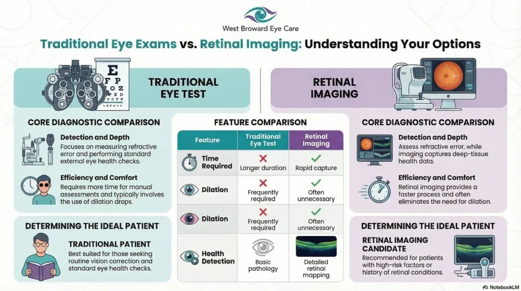
A traditional eye test measures how clearly you see using charts, lenses, and light. Retinal imaging goes further — using advanced technology to photograph and map the inside of your eye, detecting diseases like diabetic retinopathy, macular degeneration, and glaucoma before symptoms ever appear. For patients in Tamarac, FL, West Broward Eye Care offers both, combining comprehensive traditional exams with state-of-the-art Optomap and Optovue retinal imaging — all under one roof, delivered by board-certified optometric physicians with 35 years of community experience.
Most people walk into an eye exam thinking about one thing: their prescription. Can they see the board clearly? Do they need stronger glasses? But modern eye care has evolved far beyond the eye chart — and understanding the difference between a traditional eye test and retinal imaging could be one of the most important health decisions you make this year.
Whether you are a parent worried about your child’s worsening nearsightedness, a senior managing diabetes, or simply someone who wants the clearest possible picture of their long-term eye health, this guide will help you understand exactly what each type of exam offers — and which one is right for you.
What Does a Traditional Eye Test Actually Measure?
A traditional eye exam is the foundation of eye care — and for good reason. It is thorough, proven, and essential for millions of patients every year. But it is important to understand precisely what it measures, so you know where its boundaries lie.
The Visual Acuity Test (The Eye Chart)
This is the test most people picture when they think of an eye exam. You sit across the room, cover one eye, and read lines of progressively smaller letters. This measures your visual acuity — how sharp and clear your vision is at various distances. It is the starting point for every prescription for glasses or contact lenses.
Cover Tests, Color Vision & Peripheral Checks
Beyond the chart, a comprehensive traditional exam includes a series of additional evaluations. A cover test checks how well your eyes work together and detects conditions like strabismus (eye misalignment). Color vision testing identifies deficiencies that may affect daily life. Peripheral vision tests map your side vision, which is often one of the first things affected by glaucoma. Slit-lamp examinations allow the doctor to inspect the front structures of your eye — the cornea, lens, and iris — under high magnification.
What Traditional Tests Do Well — And Where They Stop
Traditional exams are highly effective at determining your prescription, detecting surface-level eye conditions, and establishing a baseline for your overall vision health. For many healthy patients under 40 with no significant risk factors, a comprehensive traditional exam may be entirely sufficient.
However, traditional testing has a fundamental limitation: it primarily evaluates the front and functional aspects of your eye. It cannot produce a detailed, high-resolution map of your retina — the light-sensitive tissue at the back of your eye that is responsible for translating what you see into signals your brain can interpret. And it is precisely in that retinal tissue where some of the most serious, vision-threatening diseases quietly take root, long before you notice any change in how you see.
What Is Retinal Imaging — And Why Is It Different?
If a traditional eye test looks at your eye, retinal imaging looks into it — deep into the structures at the back of the eye that a standard exam simply cannot reach with the same level of detail and precision.
Looking Into the Eye, Not Just At It
Retinal imaging uses specialized cameras and scanning technology to capture high-resolution images of your retina, optic nerve, and the blood vessels that nourish them. These images give your doctor a detailed, documented view of your internal eye health that can be compared year over year — allowing subtle changes to be detected long before they affect your vision or become symptomatic.
Think of it this way: a traditional eye test is like checking the exterior of a car. Retinal imaging opens the hood.
How Optomap Works: The Wide-Field Advantage
At West Broward Eye Care, the Optomap retinal imaging system captures up to 82% of your retina in a single, ultra-widefield image — without the need for dilation drops. For patients who have experienced the blur and light sensitivity that comes with traditional dilation, this is a significant comfort advantage. The scan itself takes only seconds, is completely painless, and produces a richly detailed panoramic image of your retinal health that your doctor can review with you in real time.
Traditional exams using dilation can typically view only 10–12% of the retina at a time. The difference in diagnostic coverage is significant.
How Optovue (OCT) Works: Seeing Layer by Layer
While Optomap provides the wide view, the Optovue system at West Broward Eye Care adds another critical dimension: Optical Coherence Tomography (OCT). This technology uses light waves to take cross-sectional images of your retina, allowing your doctor to examine each individual layer of retinal tissue at a near-microscopic level.
OCT is particularly powerful for detecting early-stage macular degeneration, subtle changes in the optic nerve associated with glaucoma, and fluid accumulation beneath the retinal surface — changes that are invisible to the naked eye and undetectable by standard examination alone.
| Feature | Traditional Eye Test | Retinal Imaging (Optomap/OCT) |
|---|---|---|
| Measures visual acuity | Yes | Yes |
| Detects retinal disease early | Limited | Yes |
| Requires dilation drops | Often yes | No (Optomap) |
| Views retinal layers (OCT) | No | Yes (Optovue) |
| Suitable for diabetic patients | Basic only | Highly recommended |
| Monitors myopia in children | Limited | Yes |
| Time required | 5–10 min | 5–10 min |
| Permanent image record | No | Yes |
What Conditions Can Retinal Imaging Detect That a Standard Test Cannot?
This is where retinal imaging moves from a convenience to a clinical necessity for many patients. The conditions below share a critical characteristic: they cause little to no symptoms in their early stages, meaning patients often feel fine while disease silently progresses. Early detection through retinal imaging is not just beneficial — it can be vision-saving.
Diabetic Retinopathy — Catching It Before Symptoms Appear
Diabetic retinopathy is the leading cause of blindness among working-age adults in the United States. It occurs when elevated blood sugar damages the tiny blood vessels in the retina, causing them to leak, swell, or grow abnormally. In its earliest stages, there are no visual symptoms whatsoever. A patient can have moderate diabetic retinopathy and still read the eye chart perfectly.
Optomap and Optovue imaging allow the physicians at West Broward Eye Care to detect these microscopic vascular changes at their earliest, most treatable stage — giving patients and their primary care providers the critical information they need to intervene before irreversible damage occurs.
Macular Degeneration — Monitoring the Macula Over Time
Age-related macular degeneration (AMD) affects the central portion of the retina responsible for sharp, detailed vision. Early AMD produces no pain and often no noticeable vision loss, yet structural changes in the macula are already underway. OCT imaging with Optovue can detect the earliest signs of AMD — including drusen deposits and retinal pigment changes — years before central vision is affected, enabling proactive monitoring and lifestyle interventions that can slow progression significantly.
Glaucoma — Spotting Nerve Damage at Its Earliest Stage
Glaucoma is often called the “silent thief of sight” because it destroys peripheral vision gradually and painlessly. By the time many patients notice vision loss, significant and irreversible optic nerve damage has already occurred. OCT imaging provides precise measurements of optic nerve fiber layer thickness — one of the earliest and most reliable indicators of glaucomatous damage — enabling treatment to begin at the point where it will be most effective.
Myopia Progression in Children — A Parent’s Early Warning System
For parents whose children are experiencing increasing nearsightedness, retinal imaging provides an invaluable baseline and monitoring tool. Rapidly progressive myopia in childhood is associated with a significantly elevated risk of serious retinal conditions in adulthood, including retinal detachment. Establishing a detailed retinal map early allows the team at West Broward Eye Care to track changes precisely and integrate findings into their specialized Myopia Management program.
Concerned about your retinal health or your child’s vision? The board-certified physicians at West Broward Eye Care use Optomap and Optovue technology to detect eye disease at its earliest, most treatable stage. Call or text 954-726-0204 to schedule your comprehensive exam today.
The South Florida Factor — Why Retinal Imaging Matters More Here
Geography and demographics matter in eye care — and Tamarac, FL presents a unique combination of environmental and population factors that make advanced retinal imaging not just a premium option, but a genuinely important consideration for a large portion of the local community.
High UV Exposure & Its Long-Term Impact on Retinal Health
South Florida receives some of the highest levels of ultraviolet radiation in the continental United States. Cumulative UV exposure over decades is a well-established risk factor for both macular degeneration and the development of retinal abnormalities. For patients who have spent years enjoying the Florida sunshine — as most Tamarac residents have — a comprehensive retinal baseline is a proactive and sensible investment in long-term vision health.
Tamarac’s Aging Population & the Diabetic Eye Disease Connection
Broward County has one of the largest senior populations in Florida, and Tamarac reflects that demographic profile. Older adults face elevated risks for diabetic retinopathy, macular degeneration, and glaucoma — the three leading causes of irreversible vision loss in the United States. The specialized retinal imaging capabilities at West Broward Eye Care are precisely calibrated to serve this community’s most pressing eye health needs, providing the kind of diagnostic depth that routine exams at general optical chains simply cannot match.
Why Local Families Are Choosing Advanced Screening for Their Children
The prevalence of childhood myopia is rising rapidly across the United States, and Broward County families are not exempt from this trend. Parents who are proactive about their children’s long-term vision are increasingly seeking practices that offer more than a basic prescription update. West Broward Eye Care’s combination of retinal imaging and dedicated Myopia Management services makes it the logical choice for families who want to stay ahead of their children’s eye health — not just react to it.
Traditional Eye Test vs. Retinal Imaging: A Side-by-Side Patient Guide
Understanding the clinical differences is important. But what most patients ultimately want to know is simple: which one do I actually need? The answer depends on your individual health profile, age, family history, and risk factors.
Choose a Traditional Comprehensive Exam If…
A traditional comprehensive exam may be the primary focus of your visit if you are a generally healthy adult under 40 with no personal or family history of eye disease, no systemic conditions like diabetes or hypertension, and no current visual symptoms or concerns. This exam will accurately assess your visual acuity, update your prescription, and screen for common surface-level conditions. It is a strong foundation for anyone with a low overall risk profile.
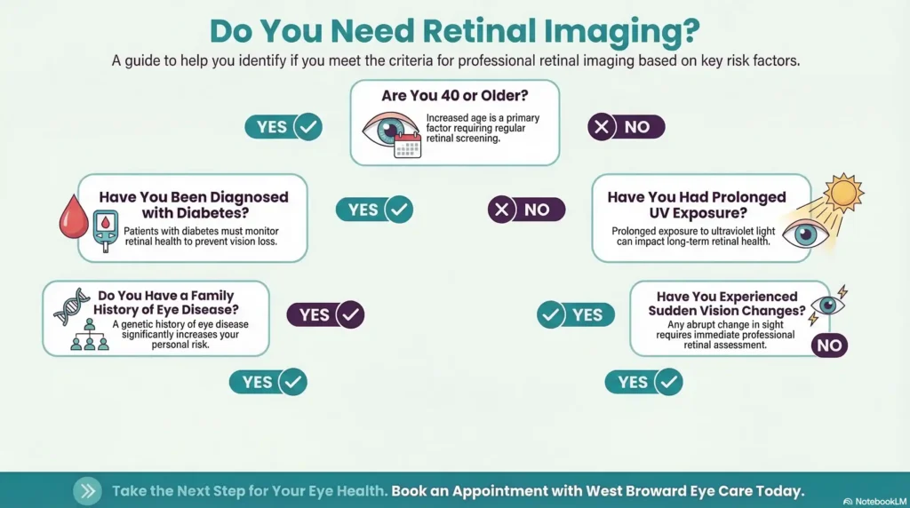
Choose Retinal Imaging If…
Retinal imaging is strongly recommended if you are 40 years of age or older, have been diagnosed with diabetes or hypertension, have a family history of glaucoma, macular degeneration, or retinal disease, have experienced sudden changes in your vision such as floaters, flashes, or blurred central vision, are a child or teenager with rapidly progressing myopia, or have a history of significant UV exposure. In South Florida, that last factor applies to the vast majority of long-term residents.
The Best Option: Why Most Tamarac Patients Benefit From Both
For most patients — and particularly for anyone over 40 or with any of the risk factors listed above — the most complete picture of eye health comes from combining a thorough traditional comprehensive exam with retinal imaging. These two approaches are not competitors; they are complements. The traditional exam establishes your visual prescription and evaluates the front structures of your eye. Retinal imaging maps the internal health of the back of your eye. Together, they give your doctor — and you — the most complete understanding of your eye health possible.
| Patient Profile | Recommended Exam Type |
|---|---|
| Healthy adult under 40, no risk factors | Comprehensive traditional exam |
| Adult 40+ or with diabetes history | Retinal imaging + comprehensive exam |
| Child with worsening nearsightedness | Myopia monitoring + retinal baseline |
| Patient with sudden vision changes | Urgent retinal imaging — same day care available |
| Senior at risk for macular degeneration | Annual Optomap + Optovue OCT |
| Post-LASIK patient | Annual retinal health monitoring |
Why West Broward Eye Care Is Tamarac’s Choice for Advanced Retinal Imaging
Choosing where to have your retinal imaging performed is not a decision to make casually. The quality of the technology, the experience of the physicians interpreting the images, and the depth of follow-up care all determine how much value that imaging truly delivers.
Optomap + Optovue: Diagnostic Technology That Sees the Full Picture
West Broward Eye Care is equipped with both the Optomap ultra-widefield retinal imaging system and the Optovue OCT platform — a combination that delivers both breadth and depth in retinal analysis. Optomap captures the panoramic view, while Optovue examines individual retinal layers at near-microscopic resolution. This dual-technology approach ensures that nothing is missed and that your physician has the most complete diagnostic picture available from a single visit.
35 Years of Trust. 885 Reasons to Choose Us.
West Broward Eye Care has been a cornerstone of eye health in the Tamarac community for 35 years. That longevity is not simply a number — it represents decades of patient relationships, generational family care, and a sustained commitment to investing in the best available technology and talent. With over 885 Google reviews reflecting consistent praise for thoroughness, warmth, and expertise, the practice’s reputation is one of the most validated in the region.
Board-Certified Care With a Neighborhood Feel
Every exam at West Broward Eye Care is performed by highly skilled, board-certified optometric physicians who combine advanced clinical training with a genuine commitment to patient-centered care. Patients consistently describe the atmosphere as warm, professional, and unhurried — a place where questions are welcomed and answers are given thoroughly. This is not a volume-driven optical chain. It is a practice built on relationships, rooted in one community, and dedicated to the long-term vision health of every patient who walks through the door.
West Broward Eye Care has been serving the Tamarac community for 35 years. Our advanced Optomap retinal imaging gives you the clearest picture of your eye health — without dilation. Visit us at 7822 N. University Dr., Tamarac, FL 33321 or call or text 954-726-0204 to book your appointment.
What to Expect During a Retinal Imaging Exam at West Broward Eye Care
One of the most common reasons patients hesitate to request retinal imaging is simple uncertainty: they do not know what the process involves. The reality is that retinal imaging at West Broward Eye Care is fast, comfortable, and entirely non-invasive.
Before Your Appointment
No special preparation is required for Optomap retinal imaging. Unlike traditional dilation, you do not need to arrange for a driver or plan for hours of light sensitivity afterward. Simply arrive for your scheduled appointment as you normally would. If you wear contact lenses, you may be asked to remove them briefly for certain components of the exam.
During the Scan — Fast, Comfortable & Dilation-Free
The Optomap scan itself takes approximately 30 seconds per eye. You will look into the device, focus on a small fixation target, and a gentle flash of light will capture the panoramic image. There is no contact with your eye, no discomfort, and no aftereffects. The Optovue OCT scan is equally fast and non-contact — you simply rest your chin on a support and look ahead while the device scans. Most patients are genuinely surprised by how quick and comfortable the entire process is.
After the Scan — Understanding Your Results With Your Doctor
This is where West Broward Eye Care truly distinguishes itself. Your images are reviewed immediately by your physician, who will walk you through what they show — explaining any findings in clear, accessible language and placing them in the context of your overall eye health and medical history. You leave not just with a prescription, but with a genuine understanding of the current state of your retinal health and any steps recommended going forward.
🔗 Local Resources & Citations
1. Florida Department of Health in Broward County (DOH-Broward) (Government — .gov) The official state health authority for Broward County publishes local chronic disease data — including diabetes and vision impairment statistics — that confirms why residents of Tamarac should prioritize annual retinal screenings.
2. FLHealthCHARTS — Florida Department of Health Vision Disability Data (Government — .gov) This official state data tool tracks vision disability rates by Florida county over ten years, giving Tamarac patients a factual, local benchmark for understanding how prevalent eye disease is in their own community.
3. Nova Southeastern University College of Optometry — Fort Lauderdale, FL (Educational — .edu) Located in Broward County, NSU’s nationally accredited optometry college conducts active clinical research in retinal disease, myopia control, and glaucoma — making it the region’s primary academic authority on the conditions that retinal imaging is designed to detect.
4. Optos — Official Optomap Ultra-Widefield Retinal Imaging Technology (Official Manufacturer — Technical Specifications) The manufacturer of the Optomap device used at West Broward Eye Care publishes full clinical specifications confirming that optomap is the only clinically validated ultra-widefield retinal image that can capture 82% or 200° of the retina in a single capture — the technical proof point behind the article’s diagnostic coverage claims.
Insurance & Cost — Is Retinal Imaging Covered?
Coverage for retinal imaging varies depending on your insurance plan and the clinical indication for the scan. In many cases, retinal imaging is covered when it is medically indicated — for example, for patients with diabetes, glaucoma risk, or macular degeneration monitoring. In other cases, it may be offered as an elective enhancement to a routine exam for a modest additional fee.
West Broward Eye Care accepts a wide range of insurance plans and is committed to providing options for all budgets. The team will review your coverage with you at the time of your visit and ensure full transparency around any out-of-pocket costs before your exam begins. For a service that provides the level of diagnostic value retinal imaging delivers, most patients find the investment — whether covered by insurance or not — well worth the peace of mind it provides.
For specific coverage questions, contact the practice directly at 954-726-0204 or email info@wbeca.com.
Conclusion — See More. Know More. Protect More.
Your vision is one of your most valuable assets — and protecting it begins with understanding it. A traditional eye test tells you how clearly you see today. Retinal imaging tells you what is happening inside your eye right now, and what may be developing long before you notice any change in your vision.
For the families, seniors, and individuals of Tamarac and the greater Broward County community, West Broward Eye Care has been the trusted answer for 35 years. With state-of-the-art Optomap and Optovue technology, board-certified physicians, and a deeply patient-centered approach, the practice offers something no national chain can replicate: the power of advanced diagnostics delivered with the warmth of a neighbor who genuinely cares about your long-term well-being.
See more. Know more. Protect more. Your eyes deserve nothing less.
Ready to see the full picture of your eye health? Schedule your advanced retinal imaging exam with the trusted experts at West Broward Eye Care. Call or text: 954-726-0204 7822 N. University Dr., Tamarac, FL 33321 Monday–Thursday 9:00 AM–5:00 PM | Friday 9:00 AM–4:00 PM
Frequently Asked Questions
-
Retinal imaging is used to photograph and map the internal structures of the eye — including the retina, optic nerve, and blood vessels — at a level of detail that standard eye exams cannot achieve. It is used to detect, monitor, and manage serious eye conditions such as diabetic retinopathy, macular degeneration, glaucoma, and retinal detachment, often before any symptoms appear. At West Broward Eye Care in Tamarac, FL, retinal imaging using Optomap and Optovue technology is a core component of comprehensive eye health evaluation for patients of all ages.


